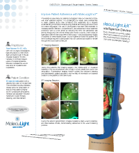
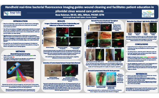
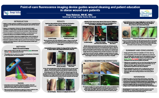
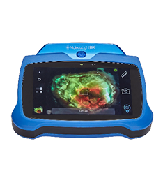
Knowledge is a key factor for patients to engage in their own care and comply with their treatment regime.1 It is challenging for health care professionals to teach patients about their conditions and treatments as it requires understanding of complex concepts using medical terminology and can involve non-native languages. The use of photographs has been demonstrated to increase patients’ attention, comprehension, and adherence to their treatment protocols,2 and is a tool aimed at reducing the 50% non-adherence rate among those living with chronic illness and chronic wounds, which costs an estimated USD$100 billion/year (€80.3 billion/year).1 Using fluorescence images of bacteria to demonstrate proper wound hygiene provided this patient with the knowledge required to participate in his own care and advocate for himself through the wound care continuum.

Clinician's Testimonial
"No one had been cleaning in this crease. I have since imaged a couple of other patients with bacteria in creases and I asked them to take a picture of the MolecuLight i:X fluorescent image on their phone and show it to their nurse, so the nurse will know where to clean. It will make a difference."
Rose Raizman, RN-EC, MSc, Scarborough & Rouge Hospital, Toronto, ON, Canada
Clinician Profile
With over 19 years of experience, Rose leads the Save Our Skin (SOS) team at Scarborough & Rouge Hospital located in Toronto, Canada, to combat pressure ulcers of hospital inpatients. She also oversees the wound care clinic for inpatients and outpatients.
Clinical Synopsis
Patient Condition: 45 year old male patient with a diabetic foot ulcer on his right heel. Comorbidities including diabetes, obesity, and prior amputation of toes, put this patient at higher risk for subsequent amputations. General care paradigm included hyperbaric oxygen therapy, cleaning, and debridement.
1st Imaging Session
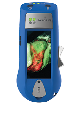
During this patient’s first imaging session, the MolecuLight i:X visualized bacteria in the wound periphery and off-site in a foot crease from a prior toe amputation. Fluorescence Imaging Mode™ guided the clinician’s cleaning and debridement, patient education, and the relay of information on bacterial location to the patient’s home care nurse.
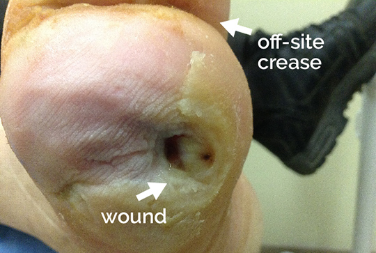 |
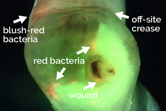 Arrows indicate regions of red and blush-red bacterial fluorescence in wound periphery and off-site |
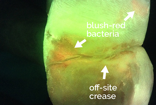 Arrows indicate regions of blush-red bacterial fluorescence off-site and in crease |
2nd Imaging Session
During the second appointment, FL-images revealed a clean wound periphery and less bacteria off-site, demonstrating adherence to the wound cleaning protocol.
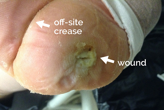 Image of wound and off-site crease at 2nd imaging session |
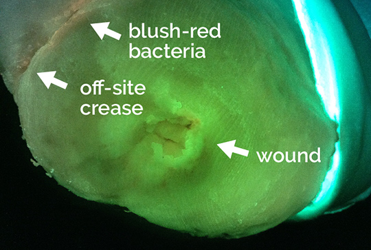 Arrow indicates reduction of blush-red bacterial fluorescence in off-site crease |
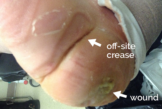 Image of off-site crease |
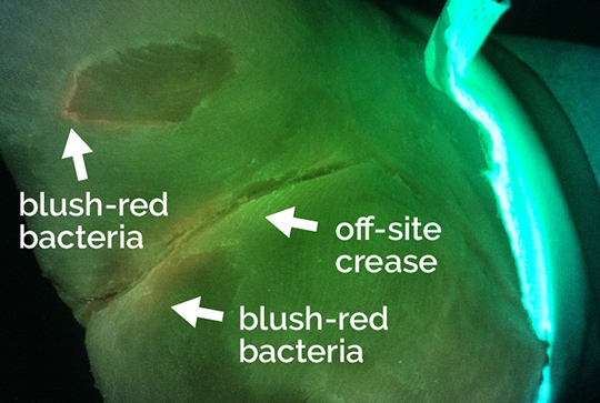 Arrows indicate reduction of off-site blush-red bacterial fluorescence |
Patient Testimonial
“This device is very useful because it creates some knowledge, so you can see what’s actually going on. I have some of the images stored on my phone so that I can show them to my CCAC home care nurse who does the dressing changes so he gets to see as well. It would be useful in CCAC practice because it would relieve the visits coming to see the surgeon or primary wound care specialists. Conveyance in wound care is critical because the wound care nurse needs to convey what is going on to the CCAC person doing the dressing changes. This device helps convey that information, so they can actually see where the problem areas are and where to focus on.”
At a Glance
| Wound etiology & location | Diabetic foot ulcer, right toe |
| Patient demographics | Male, 45 years old |
| Patient-related challenges | Diabetes
Overweight/poor diet Prior amputation of toes |
| Patient’s general care paradigm | Cleaning and debridement
Hyperbaric oxygen therapy |
| Clinician stated utility of the MolecuLight i:X | Detection of off-site bacteria
Guided clinician cleaning and debridement Patient education & adherence to at-home cleaning protocol |
