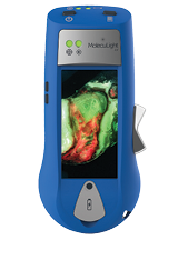
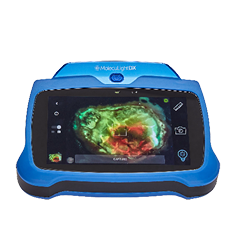
How does the MolecuLight i:X work?
In a darkened room, or using a DarkDrape™, the MolecuLight i:X shines a safe violet excitation light (405 nm) on a wound. This causes wound components (skin, slough, blood, bacteria, etc.) to fluoresce (or emit light) in different colours1–4.The i:X device displays and captures images of the most informative of these fluorescent colours. Green fluorescence from skin components (i.e. collagen and fibrin) provides anatomical context. Red and cyan fluorescence are associated with regions of bacterial at loads of >104 CFU/g1–3, which is typically moderate-to heavy growth1–3, as demonstrated in multiple clinical studies. Bacterial loads of less than 104 CFU/g, such as the loads of commensal bacteria on healthy skin, are not detected by the MolecuLight i:X1,4.
In addition to bacterial detection, the i:X also performs digital wound area measurement to document and longitudinally monitor the progress of wounds.
Which types of bacteria can be detected?
Pre-clinical and clinical studies have shown that the MolecuLight i:X can detect red fluorescence from Gram positive, Gram negative, aerobic and anaerobic bacterial species1,5.
Pre-clinical research has demonstrated the following species can produce red fluorescence detectable by the MolecuLight i:X in vitro5 (see list below). However, there are many bacterial species not tested here that may also produce red fluorescence6.
Gram negative aerobic species
- Pseudomonas aeruginosa
- Escherichia coli
- Proteus mirabilis
- Proteus vulgaris
- Enterobacter cloacae
- Serratia marcescens
- Acinetobacter baumannii
- Klebsiella pneumoniae
- Klebsiella oxytoca
- Morganella morganii
- Stenotrophomonas maltophilia
- Citrobacter koseri
- Citrobacter freundii
- Aeromonas hydrophilia
- Alcaligenes faecalis
- Pseudomonas putida
Gram positive aerobic species
- Staphylococcus aureus
- Staphylococcus epidermis
- Staphlyoccus lugdunensis
- Staphylococcus capitis
- Corynebacterium striatum
- Bacillus cereus
- Listeria monocytogenes
Anaerobic species
- Bacteroides fragilis
- Clostridium perfringens
- Peptostreptococcus anaerobius
- Propionibacterium acnes
- Veillonella parvula
How easy is the MolecuLight i:X to use?
The MolecuLight i:X device is straight-forward to learn and operate. The device’s user interface is designed to be intuitive and easy to use, without requiring a technical background. The device is hand-held and does not require any contrast agents.
MolecuLight provides comprehensive on-site training for all staff and there is a library of online e-learning modules, online assessments, and videos to assist all wound care practitioners with image interpretation.
Why does fluorescence imaging (with the i:X device) need to be performed in the dark?
In order for the i:X to visualize fluorescence in wounds indicative of elevated bacterial burden (>104 CFU/g), fluorescence imaging using the MolecuLight i:X requires a dark room. Darkness is required so that fluorescence signals emitted from the bacteria (and other biological components) in a wound are clearly visualized by the device. In order to ensure a room is sufficiently dark, the i:X has an ambient light sensor to inform the user if the room is dark enough.
If a clinical room can’t be darkened (e.g. an open ward or a treatment room with large windows), a DarkDrape® is available as a disposable, portable dark environment to provide the required darkness for successful point-of-care fluorescence imaging.
Insufficient darkness may lead to images that are not able to be accurately interpreted.
What does it mean that the device shows ‘real-time’ data?
When the “fluorescence mode” (FL-mode) is activated in a dark environment, and an image or video is taken, red or cyan signals show up immediately on the i:X screen. When red or cyan fluorescence is present, this indicates an increased likelihood that the wound contains elevated bacterial load. No data processing or contrast agents are required. Visualization of fluorescence is instantaneous as long as fluorescence mode is on and the room is dark. As such, the wound care practitioner can move the device around the patient’s wounds to improve the detection of elevated bacterial burden (104 CFU/g) over CSS alone.
Is the violet light of the i:X device safe?
Yes. The i:X device illuminates with violet light, which is safe, and not with ultraviolet (UV) light. The i:X device is classified in Risk Group 1 and the laser is Class 1. This means there is not sufficient energy produced by the device to damage skin or eyes in normal use. No safety eye wear is required. However, it is advised to not point the device towards the eyes.
Can I miss the fluorescence signal if there is blood in the wound?
Blood does not fluoresce. It preferentially absorbs the excitation light thereby reducing the likelihood of (bacteria/tissue) fluorescence being generated. Bacteria covered by surface blood may be missed during fluorescence imaging, but this is rare as users should remove blood during wound assessment (when possible). It is recommended that fluorescence imaging be performed after (surface) blood has been removed from the wound bed and periwound areas.
Can images be transferred off the i:X device?
Yes. Images, measurements and videos can be transferred from the i:X device to any computer or network using the i:X Connecting Cable or via local Wi-Fi.
Does anything other than bacteria fluoresce?
Yes, the MolecuLight i:X sees all fluorescence. Non-biological sources such as bed sheets, tattoo inks and fluorescent dyes can also fluorescence and may be detected by the MolecuLight i:X. It is always important to compare fluorescence images to standard images for the most accurate interpretation. See this image interpretation publication.
If you have additional questions, please contact us.
