Home
Transform Wound Management with Multimodal Imaging
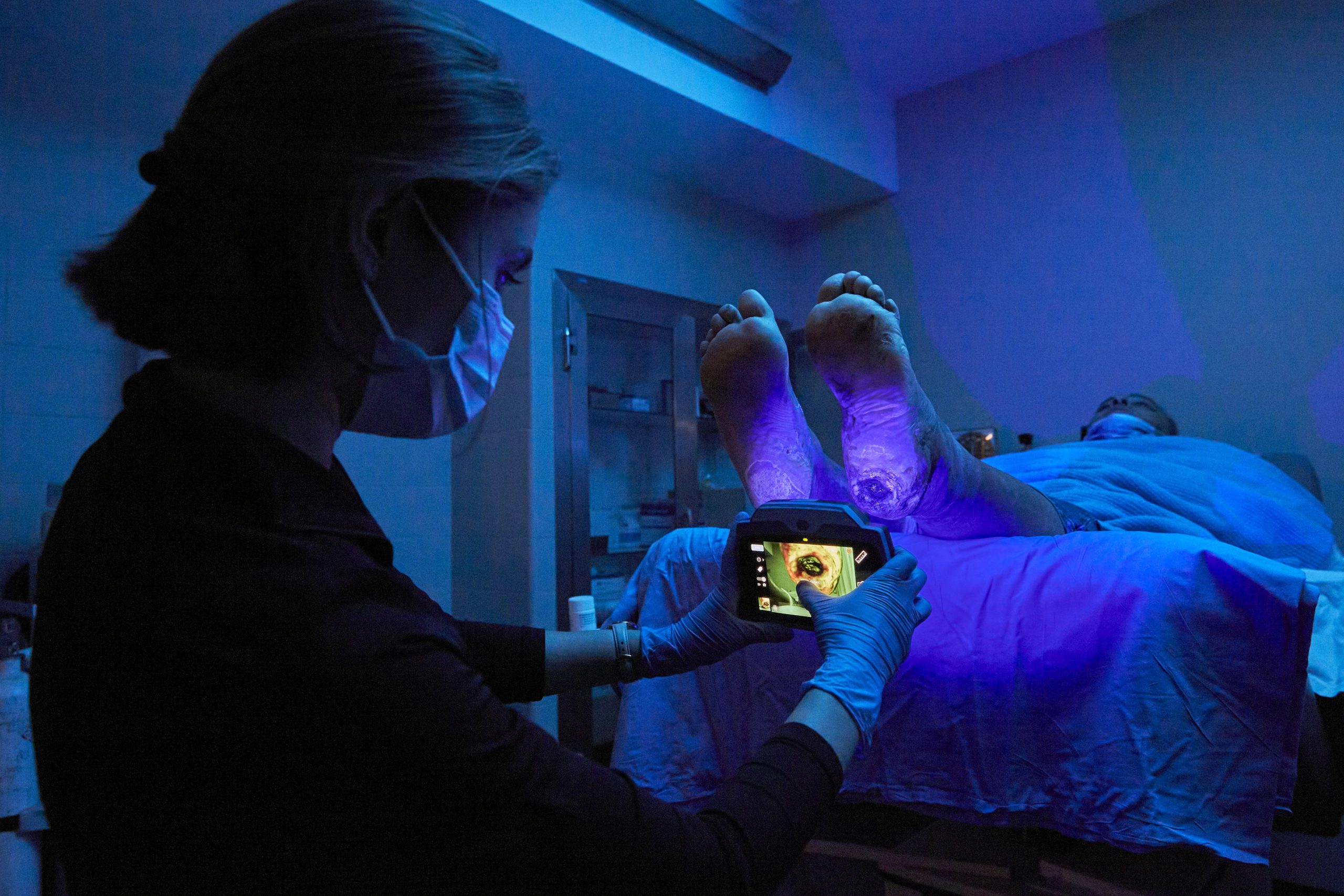
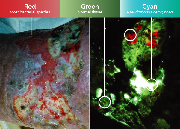
Detect
Elevated Bioburden
Locate in real-time regions containing high bacterial loads with the only non-invasive, contrast-free, FDA-cleared Class II point of care fluorescence wound imaging device.1,2
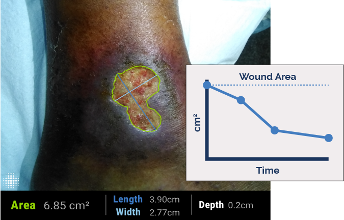
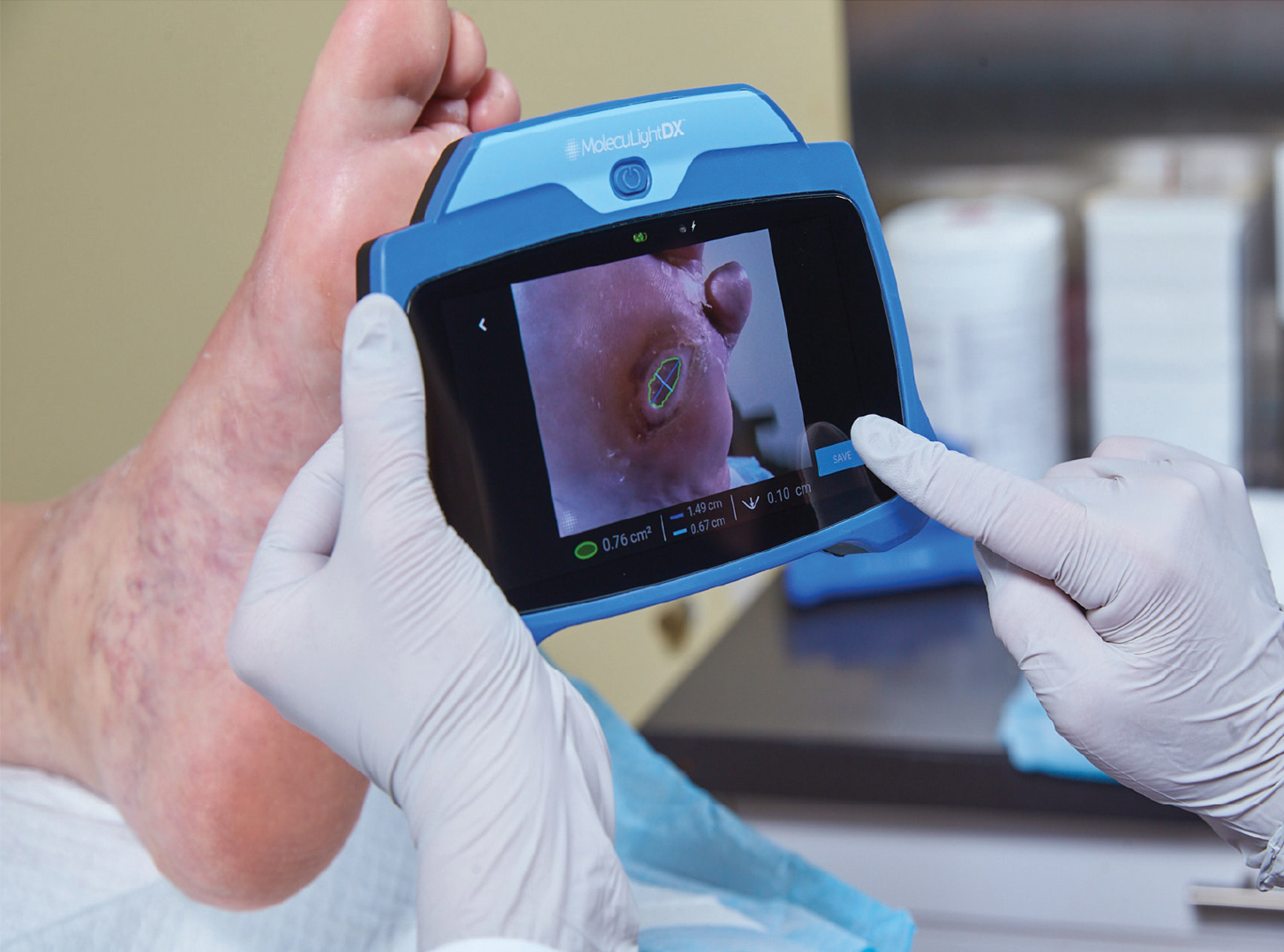
Measure
Wounds Accurately
Industry-leading validated digital wound measurement in an instant - no stickers or patient contact required. Measurements are automatically monitored and tracked over time to see wound progression.
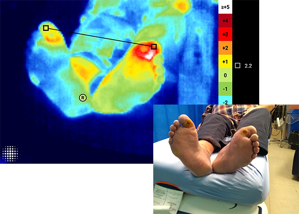
See
Thermal Changes
Quantify and document skin temperature differences with ±0.5°C accuracy. Visualize variations with a dynamic thermal map.
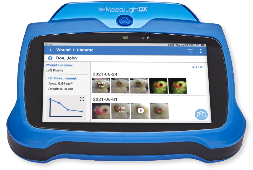
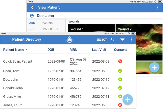
Document
With Ease
Effortless tracking and storage of wound images and details. This comprehensive evidence-based documentation helps to monitor changes in wound bioburden, treatment effectiveness and wound healing over time.

Wound Area Reduction
A prospective study showed 79% reduction in wound area by 12 weeks, when harmful bacteria detected by MolecuLight are removed.1

Wound Healing
A randomized controlled trial demonstrated 2x more wounds healed by 12 weeks using MolecuLight to inform wound care vs. Standard of Care.3

Equitable Care
In a clinical trial MolecuLight significantly improved detection of high bacterial loads in wounds across all skin tones over Standard of Care.4

Enhanced Imaging Platform
Class II Device
Fluorescence wound imaging device validated for detection of wounds with bacteria >104 CFU/g.5
EHR Integrated
Integrated with all leading EHRs
Secure Platform
High security measures HIPAA, SOC II Type 2
Reimbursed
Only billable fluorescence imaging device for wound care


