Clinical Image Gallery
Below are a series of images/videos highlighting how the MolecuLight i:X helped clinicians detect elevated bacterial burden (>104 CFU/g) in wounds and digitally measure wounds.
Media List
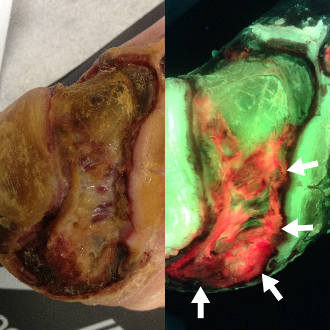
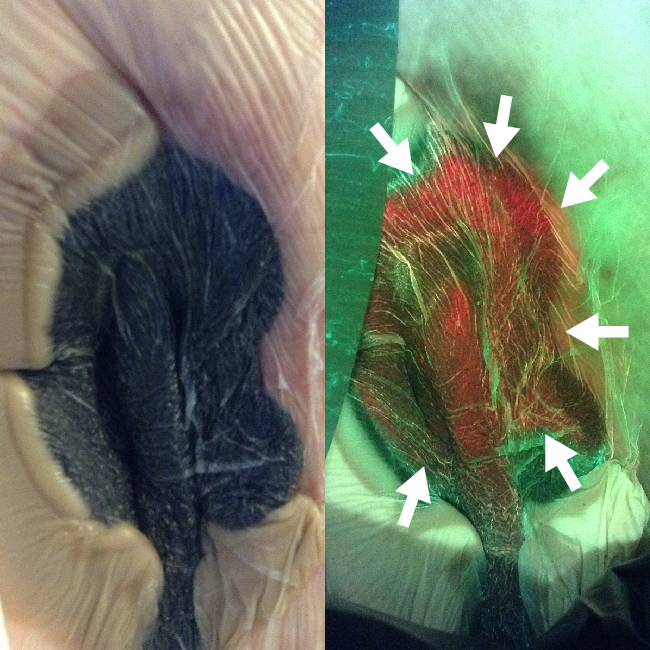
Diabetic Foot Ulcer, Heel
IMAGE
Patient sought treatment for a large diabetic foot ulcer that developed after one day spent
Figure 1: ST-image
Figure 2: FL-image
Diabetic Foot Ulcer, Heel
Patient sought treatment for a large diabetic foot ulcer that developed after one day spent wearing a pair of high heels.

Anatomical Location:
Right Foot, Heel
Image/Video Provided By:
Rose Raizman, RN-EC, MSc, Scarborough & Rouge Hospital, Toronto, ON, Canada
Case ID:
MolecuLight Clinical Case 0040

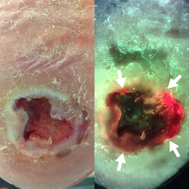
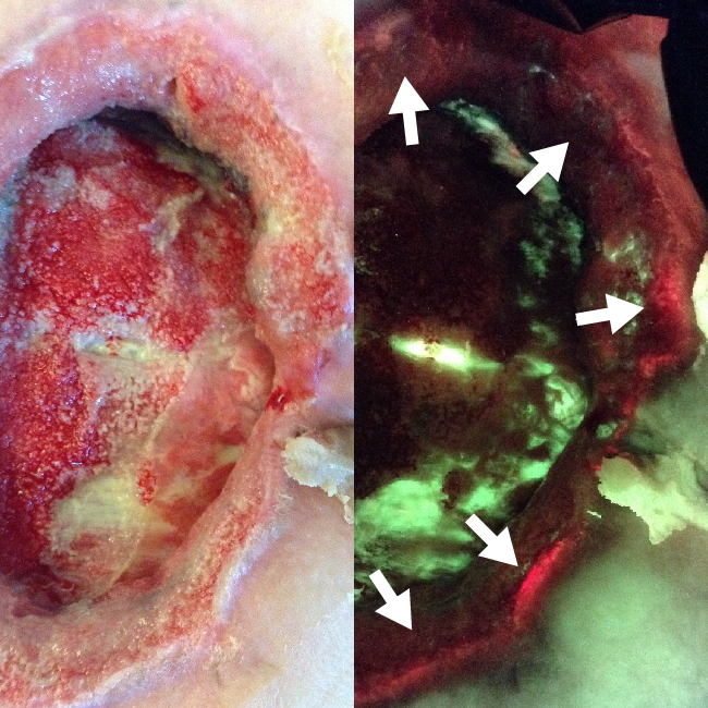
Diabetic Foot Ulcer, Toe
IMAGE
Images taken after initial debridement; wound further debrided under MolecuLight i:X guidance until red (bacterial)
Figure 1: ST-image
Figure 2: FL-image
Diabetic Foot Ulcer, Toe
Images taken after initial debridement; wound further debrided under MolecuLight i:X guidance until red (bacterial) fluorescence signal no longer detected.

Anatomical Location:
Left Foot, Toe
Image/Video Provided By:
Rose Raizman, RN-EC, MSc, Scarborough & Rouge Hospital, Toronto, ON, Canada
Case ID:
MolecuLight Clinical Case 0045
Diabetic Foot Ulcer, Heel
IMAGE
Patient presented with deteriorating wound despite aggressive offloading and good vascularity; FL-image revealed significant bioburden
Figure 1: ST-image
Figure 2: FL-image
Diabetic Foot Ulcer, Heel
Patient presented with deteriorating wound despite aggressive offloading and good vascularity; FL-image revealed significant bioburden (red) after routine cleaning and debridement which prompted clinician to switch to an antimicrobial dressing.

Anatomical Location:
Left Foot, Heel
Image/Video Provided By:
David Russell, MD, Leeds General Infirmary, Leeds, UK
Case ID:
MolecuLight Clinical Case 0056
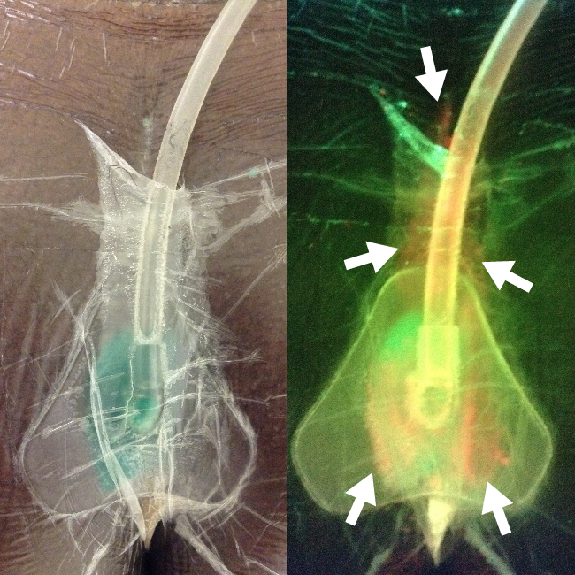
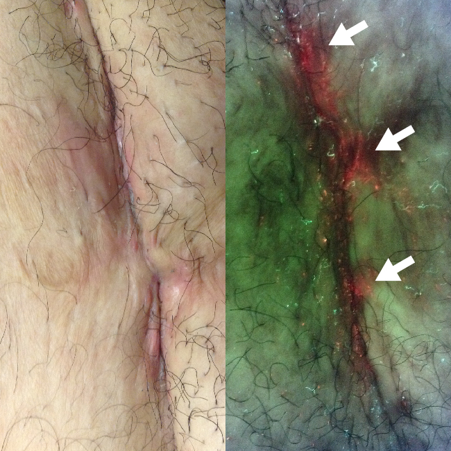
Pilonidal Sinus
IMAGE
FL-image shows extraction of bacteria via a negative pressure vacuum pump under the sealed wound
Figure 1: ST-image
Figure 2: FL-image
Pilonidal Sinus
FL-image shows extraction of bacteria via a negative pressure vacuum pump under the sealed wound dressing.
Anatomical Location:
Pilonidal Sinus
Image/Video Provided By:
Rose Raizman, RN-EC, MSc, Scarborough & Rouge Hospital, Toronto, ON, Canada
Case ID:
MolecuLight Clinical Case 0019
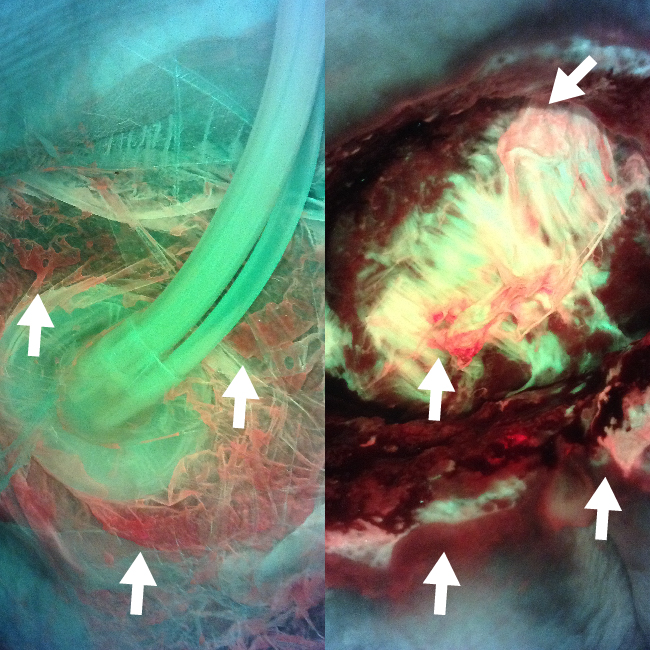
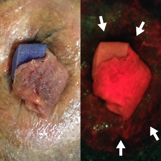
Pressure Ulcer, Sacrum
IMAGE
FL-image shows extraction of bacteria via a negative pressure vacuum pump under the sealed wound
Figure 1: FL-image
Figure 2: FL-image
Pressure Ulcer, Sacrum
FL-image shows extraction of bacteria via a negative pressure vacuum pump under the sealed wound dressing.

Anatomical Location:
Sacrum
Microbiology Results:
Swabs confirmed heavy growth of E. coli, Enterococcus faecalis, and Staphylococcus aureus
Image/Video Provided By:
Rose Raizman, RN-EC, MSc, Scarborough & Rouge Hospital, Toronto, ON, Canada
Case ID:
MolecuLight Clinical Case 0034
Pressure Ulcer, Sacrum
IMAGE
Negative pressure wound therapy on sacral pressure ulcer; FL-image revealed widespread bacteria prompting an early
Figure 1: ST-image
Figure 2: FL-image
Pressure Ulcer, Sacrum
Negative pressure wound therapy on sacral pressure ulcer; FL-image revealed widespread bacteria prompting an early dressing change and re-evaluation of the patient’s treatment plan.
Anatomical Location:
Sacrum
Microbiology Results:
Swabs confirmed heavy growth of E. coli, Enterococcus faecalis, and Staphylococcus aureus
Image/Video Provided By:
Rose Raizman, RN-EC, MSc, Scarborough & Rouge Hospital, Toronto, ON, Canada
Case ID:
MolecuLight Clinical Case 0041
Pressure Ulcer, Sacrum
IMAGE
FL-image revealed widespread bacteria prompting an early dressing change and switch to an instillation negative
Figure 1: ST-image
Figure 2: FL-image
Pressure Ulcer, Sacrum
FL-image revealed widespread bacteria prompting an early dressing change and switch to an instillation negative pressure wound therapy device.
Anatomical Location:
Sacrum
Microbiology Results:
Swabs confirmed heavy growth of E. coli, Enterococcus faecalis, and Staphylococcus aureus
Image/Video Provided By:
Rose Raizman, RN-EC, MSc, Scarborough & Rouge Hospital, Toronto, ON, Canada
Case ID:
MolecuLight Clinical Case 0041
Surgical Site Infection, C-section
IMAGE
FL-image revealed red fluorescing bacteria in skin fold at the C-section surgical site; Clinician used
Figure 1: ST-image
Figure 2: FL-image
Surgical Site Infection, C-section
FL-image revealed red fluorescing bacteria in skin fold at the C-section surgical site; Clinician used images to guide cleaning and patient education about at-home cleaning.

Anatomical Location:
Lower Abdomen
Image/Video Provided By:
Rose Raizman, RN-EC, MSc, Scarborough & Rouge Hospital, Toronto, ON, Canada
Case ID:
MolecuLight Clinical Case 0003
Surgical Site Infection, Hernia Repair
IMAGE
Images taken 1 year post-surgery; FL-image revealed bacteria in the wound periphery outside the region
Figure 1: ST-image
Figure 2: FL-image
Surgical Site Infection, Hernia Repair
Images taken 1 year post-surgery; FL-image revealed bacteria in the wound periphery outside the region which was routinely being cleaned; Clinician used images to guide thorough cleaning and apply larger antimicrobial dressing to cover the entire region of contamination.
Anatomical Location:
Abdomen, close to umbilicus
Image/Video Provided By:
Rose Raizman, RN-EC, MSc, Scarborough & Rouge Hospital, Toronto, ON, Canada
Case ID:
MolecuLight Clinical Case 0009


